| Home | Frontal | Maxillary | Ethmoid: Normal | Ethmoid: Abnormal | Sphenoid | Interactive Atlas | Quiz |
Ethmoid Sinus: Inflammatory Sinus Disease and Sequela |
|
Ethmoid inflammatory sinus disease can involve either the anterior or posterior ethmoid sinuses which have separate drainage pathways. Recall that the basal lamellae of the middle turbinate anatomically separates the ethmoid sinuses into the anterior ethmoid which drains into middle meatus and posterior ethmoid which drains into the sphenoethmoidal recess and superior meatus. As a result of the dual drainage pathways ethmoid sinus disease can also be a part of a spectrum of inflammatory sinus disease related to inflammatory disease of the ostiomeatal unit, infundibulum and spheoethmoidal recess. Ethmoid sinus disease can less commonly result in mucoceles, but due to the thin lamina papyracea and valveless ethmoidal veins can occasionally result in orbital extension of disease and cavernous sinus thrombosis.
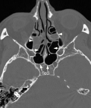
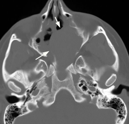
Axial image with arrowheads pointing to anterior ethmoid sinus disease. Long arrows point to clear posterior ethmoid air cells. Short arrows point to clear sphenoid sinus with (*) marking the sphenoid sinus ostium.
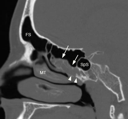
Sagittal image with arrows pointing to posterior ethmoid sinus disease and arrowheads showing involvement of the ostium and sphenoethmidal recess. (SpS: sphenoid sinus, FS: frontal sinus, MT: middle turbinate)
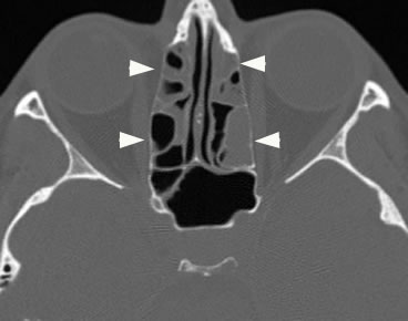
Axial image with arrowheads pointing to both anterior and posterior ethmoid sinus disease.
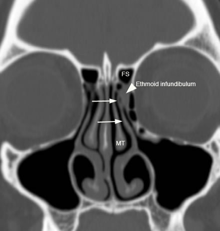
Coronal image with arrowhead pointing to ethmoid infundibulum sinus disease. In this patient the arrows are pointing to the uncinate process which connects to the skull base so the inferior compartment of the frontal sinus drainage pathway (FSDP) is the ethmoid infundibulum. (FS: frontal sinus, MT: middle turbinate)
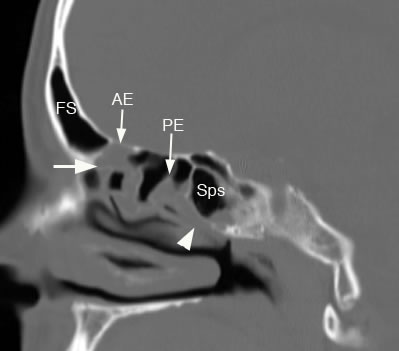
Sagittal image demonstrates more extensive involvement of the paranasal sinuses including frontal sinus (FS) and frontal sinus drainage pathway (large arow), both anterior ethmoid (AE) and posterior ethmoid sinuses (PE) marked by arrows, and sphenoid sinus (SpS) with arrowhead pointing to sphenoid sinus ostium and sphenoethmoidal recess.
Axial image with arrows pointing to expansile ethmoid mucocele.
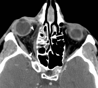
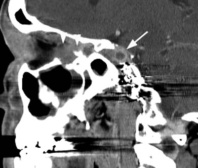
Axial post-contrast image with arrowheads pointing to orbital extension of ethmoid sinus disease marked by arrow.
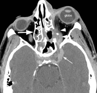
Axial post-contrast image demonstrates ethmoid sinus disease (large arrow) with orbital extension (arrowheads) resulting in cavernous sinus thrombosis (small arrow) and proptosis of left globe. Heterogeneous low density material within the enlarged cavernous sinus represents thrombus.
Sagittal post-contrast image again demonstrating the cavernous sinus thrombosis with clot (arrow) and orbital extension of disease marked by the arrowheads.
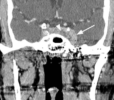
Coronal post-contrast image with arrow demonstrating the enlarged cavernous sinus due to sinus thrombosis with clot (arrowhead).
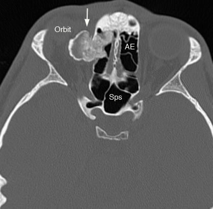
Axial image with arrow pointing to benign osteoma arising from the anterior ethmoid sinuses (AE) with adjacent orbital extension resulting in proptosis. Osteomas can result in narrowing and obstruction of the infundibulum and middle meatus. They are most common in the frontal sinuses. (SpS: sphenoid sinus)
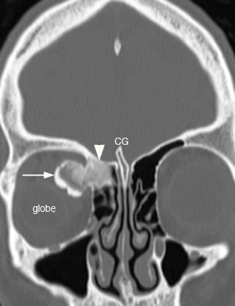
Coronal image with arrow pointing to the ethmoid osteoma extending into orbit and arrowhead showing involvement of the fovea ethmoidalis.(CG: crista galli) |