| Home | Frontal | Maxillary: Normal | Maxillary: Abnormal | Ethmoid | Sphenoid | Interactive Atlas | Quiz |
Maxillary Sinus: Inflammatory Sinus Disease and Sequela |
|
Maxillary sinus mucociliary drainage flows through the sinus ostium into the infundibulum which joins the hiatus semilunaris and drains into the middle meatus. The middle meatus is also the final drainage for the frontal and anterior ethmoid sinuses. The anterior ostiomeatal unit comprises the frontal sinus ostium, frontal recess, maxillary sinus ostium infundibulum and middle meatus. Therefore, a relatively common pattern of inflammatory sinus disease involves the anterior ostiomeatal unit. Acute sinus disease may be associated with air-fluid levels which if present commonly occur in the maxillary sinuses. However, it is important to remember that many patients with acute sinusitis will not have air-fluid levels. Acute sinusitis can also have a "bubbly or foamy" appearance. Rarely acute sinus disease can be aggressive with bony erosion. Mucus retention cysts are commonly seen and less commonly polyps and antrochoanal polyps.
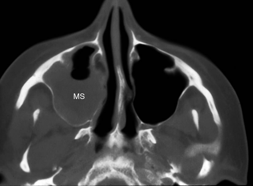
Axial image showing mucosal thicknening and an air-fluid level in the maxillary sinus (MS).
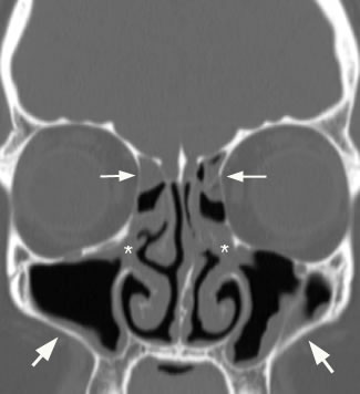
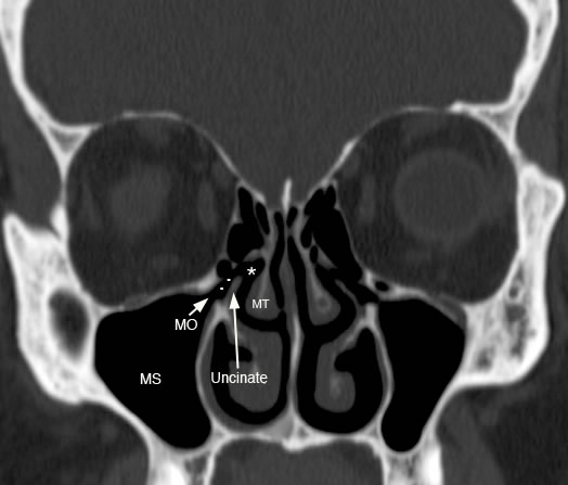
Coronal image with (*) showing obstruction of the infundibulum and on left side involvement of the hiatus semilunaris. Small arrows also demonstrate sinus disease of the anterior ethmoid air cells and larger arrows point to bilateral maxillary sinus mucosal thickening. Pattern of sinus disease involves the anterior ostiomeatal unit.
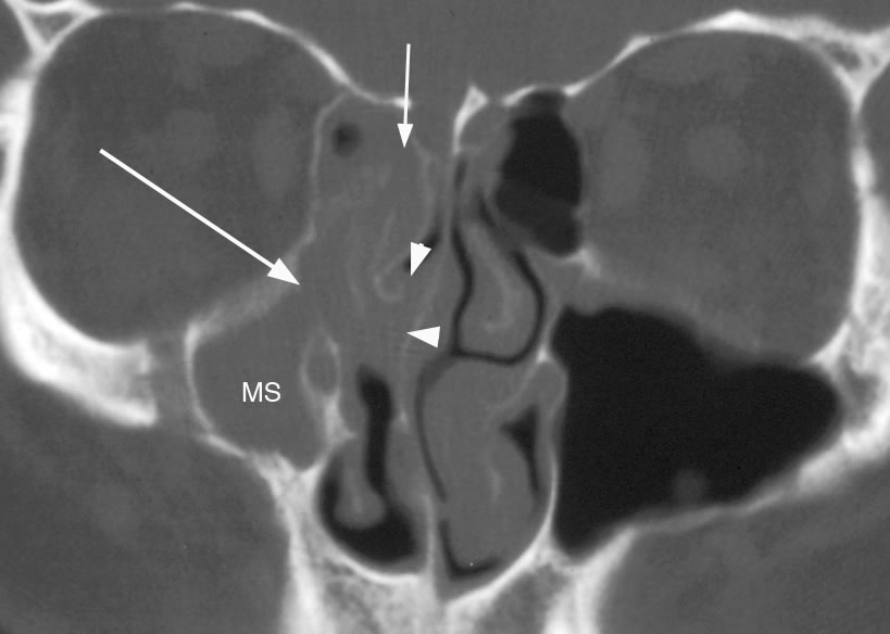
Coronal image demonstrating more extensive pattern of anterior ostiomeatal unit sinus disease with short arrow pointing to frontal recess, long arrow pointing to maxillary sinus ostium and infundibulum region with arrowheads marking the area of the hiatus semilunaris and middle meatus. Right maxillary sinus (MS) is hypoplastic.
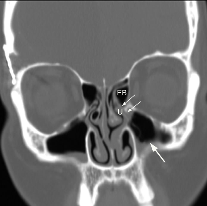
Coronal image with small arrows illustrating involvement of the maxillary sinus ostium and infundibulum and large arrow pointing to maxillary sinus mucosal thickening. (U: uncinate, EB: ethmoid bulla)
Coronal image with arrows showing involvement of the anterior ethmoid air cells. Note the right maxillary sinus mucus retention cyst and paradoxical curvature of right middle turbinate.
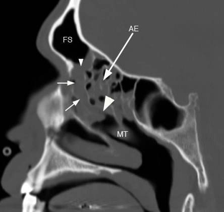
Sagittal image with small arrowhead pointing to frontal sinus ostium, short arrows showing frontal sinus drainage involvement and large arrowhead showing involvement of hitus semilunaris again demonstrating anterior ostiomeatal pattern of disease. There is also involvement of the anterior ethmoid sinus (AE). (FS: frontal sinus, MT: middle turbinate)
Axial image with arrow pointing to air-fluid level in maxillary sinus in acute sinusitis. Note the slightly bubbly appearing fluid.
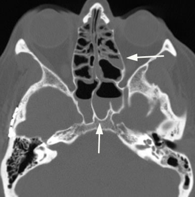
Axial image demonstrating additional case of acute sinusitis with arrows pointing to air-fluid levels in the ethmoid and sphenoid sinuses.
Coronal image with large arrow pointing to "foamy" maxillary sinus disease.
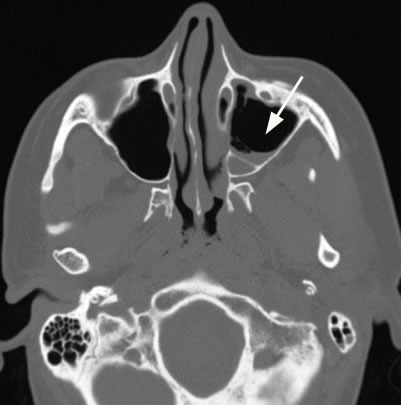
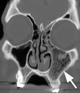
Axial image demonstrating air-fluid level in left maxillary sinus with arrow pointing to bony erosion of the posterior maxillary sinus wall in a case of acute sinusitis.
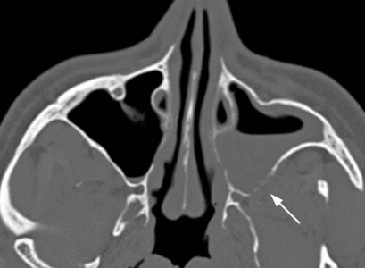
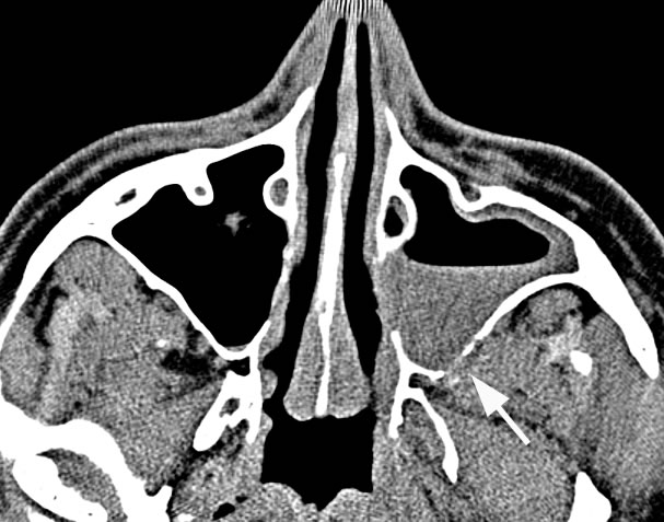
Axial image in soft tissue window with arrow pointing to posterior extension of sinus disease through the posterior maxillary sinus wall into the retroantral fat pad.
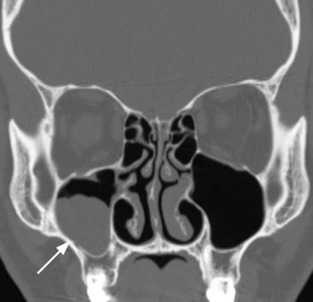
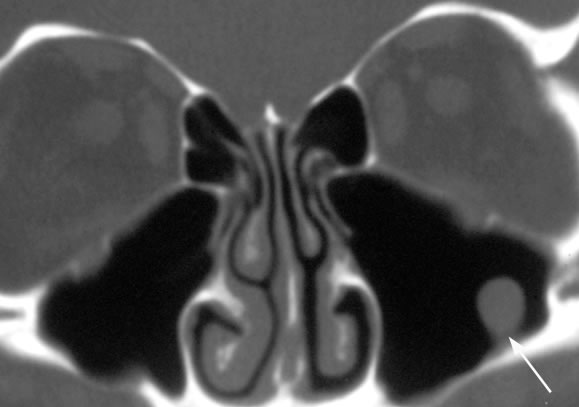
Coronal image with arrow pointing to right maxillary sinus mucus retention cyst (MRC). Notice how sinus air partially surrounds the MRC in contrast to a mucocele which completely fills and expands the sinus.
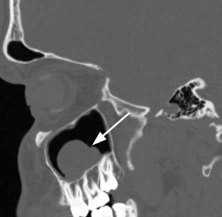
Sagittal image with arrow pointing to maxillary sinus mucus retention cyst.
Coronal image with arrow pointing to maxillary sinus polyp. Often on imaging a polyp and mucus retention cyst cannot be differentiated, but is usually of little clinical consequence.
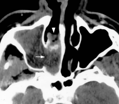
Axial image demonstrates an antrochoanal polyp that completely fills the maxillary sinus with arrow pointing to widened infundibular region with extension into the middle meatus and nasal cavity. |