
TMJ Anatomy
UW Radiology Home
TMJ Tutorial
& Function
Diagnostic Radiology Anatomy Modules
TMJ Anatomy & Function
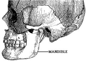
TMJ Anatomy
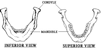
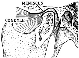
The temporomandibular joint, or TMJ, is the articulation between the condyle of the mandible and the squamous portion of the temporal bone.
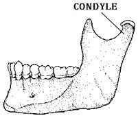
The condyle is elliptically shaped with its long axis oriented mediolaterally.
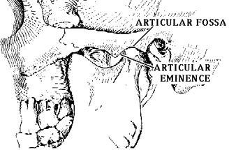
The articular surface of the temporal bone is composed of the concave articular fossa and the convex articular eminence.
The MENISCUS is a fibrous, saddle shaped structure that separates the condyle and the temporal bone. The meniscus varies in thickness: the thinner, central intermediate zone separates thicker portions called the anterior band and the posterior band. Posteriorly, the meniscus is contiguous with the posterior attachment tissues called the bilaminar zone. The bilaminar zone is a vascular, innervated tissue that plays an important role in allowing the condyle to move foreward. The meniscus and its attachments divide the joint into superior and inferior spaces. The superior joint space is bounded above by the articular fossa and the articular eminence. The inferior joint space is bounded below by the condyle. Both joint spaces have small capacities, generally 1cc or less.
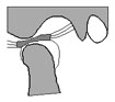
Normal TMJ Function
When the mouth opens, two distinct motions occur at the joint. The first motion is rotation around a horizontal axis through the condylar heads. The second motion is translation. The condyle and meniscus move together anteriorly beneath the articular eminence. In the closed mouth position, the thick posterior band of the meniscus lies immediately above the condyle. As the condyle translates forward, the thinner intermediate zone of the meniscus becomes the articulating surface between the condyle and the articular eminence. When the mouth is fully open, the condyle may lie beneath the anterior band of the meniscus.
 QuickTime movie of Normal TMJ motion (37K) |
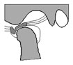
TMJ Dysfunction
Internal derangement of the TMJ is present when the posterior band of the meniscus is anteriorly displaced in front of the condyle. As the meniscus translates anteriorly, the posterior band remains in front of the condyle and the bilaminar zone becomes abnormally stretched and attenuated. Often the displaced posterior band will return to its normal position when the condyle reaches a certain point. This is termed anterior displacement with reduction.
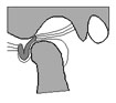
When the meniscus reduces the patient often feels a pop or click in the joint. In some patients the meniscus remains anteriorly displaced at full mouth opening. This is termed anterior displacement without reduction. Patients with anterior displacement without reduction often cannot fully open their mouths'. Sometimes there is a tear or perforation of the meniscus. Grinding noises in the joint are often present.
| QuickTime movies of Anteriorly Displaced Meniscus |
|
 With Reduction (39K) |
 Without Reduction (31K) |
For further information, contact Thurman Gillespy III, M.D.
© 1994 University of Washington Department of Radiology
All rights reserved. Do not use without written permission.
Last update: Thursday, May 10, 2001 at 2:39:43 PM.