
TMJ
UW Radiology Home
TMJ Tutorial
Arthrography
Diagnostic Radiology Anatomy Modules
TMJ Arthrography
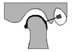
A 25 or 23 gauge needle is placed into the inferior joint space immediately posterior to the condyle. Small amounts of iodinated contrast are injected under fluoroscopy. The contrast tracks along the posterior, superior and anterior portions of the condyle. The anterior collection of contrast, called the anterior recess, normally has a smooth, tear-drop shape.
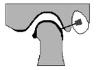
If the meniscus is perforated, contrast flows into both the superior and inferior joint recesses. However, the arthrographic needle can inadvertently puncture the meniscus and cause iatrogenic filling of both joint spaces.
| QuickTime movies of Joint Injections |
|
 Normal Inferior Joint Space (11K) |
 Perforated Meniscus (13K) |
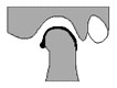
As the condyle translates anteriorly, the contrast usually empties from the anterior recess and flows posteriorly.
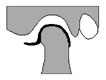
When the meniscus is anteriorly displaced, the anterior recess becomes abnormally elongated. Often the displaced meniscus is deformed or buckled, which results in a mass effect against the contrast in the anterior recess. As the condyle translates anteriorly, the mass effect against the anterior recess often increases. When the meniscus reduces, the anterior recess returns to a normal appearance.
If the meniscus does not reduce, the anterior recess remains deformed in the fully open mouth position.
| QuickTime movies of TMJ Arthrogram |
|
 Normal Joint (30K) |
 Anteriorly Displaced Meniscus with Reduction (32K) |
For further information, contact Thurman Gillespy III, M.D.
© 1994 University of Washington Department of Radiology
All rights reserved. Do not use without written permission.
Last update: Thursday, May 10, 2001 at 2:41:01 PM.